By Kat Procyk
MRI screenings are very successful at detecting breast cancer at an early stage. However, many patients struggle with claustrophobia and find it difficult to lie inside the MRI tunnel. Others may have pacemakers or metal implants that prevent them from undergoing the scan. Additionally, getting access to an MRI can often be challenging.
Cancer researchers, like Wendie Berg, Dr. Bernard F. Fisher Breast Cancer Research Professor, University of Pittsburgh School of Medicine, are exploring contrast-enhanced mammography (CEM) as a potential lower-cost alternative to MRI screenings. CEM has a detection rate similar to that of MRI screenings, but it has a far shorter examination time and makes it simpler to catch cancer earlier in patients with dense breast tissue, a condition in which breasts have a higher proportion of fibrous and glandular tissue compared to fatty tissue.
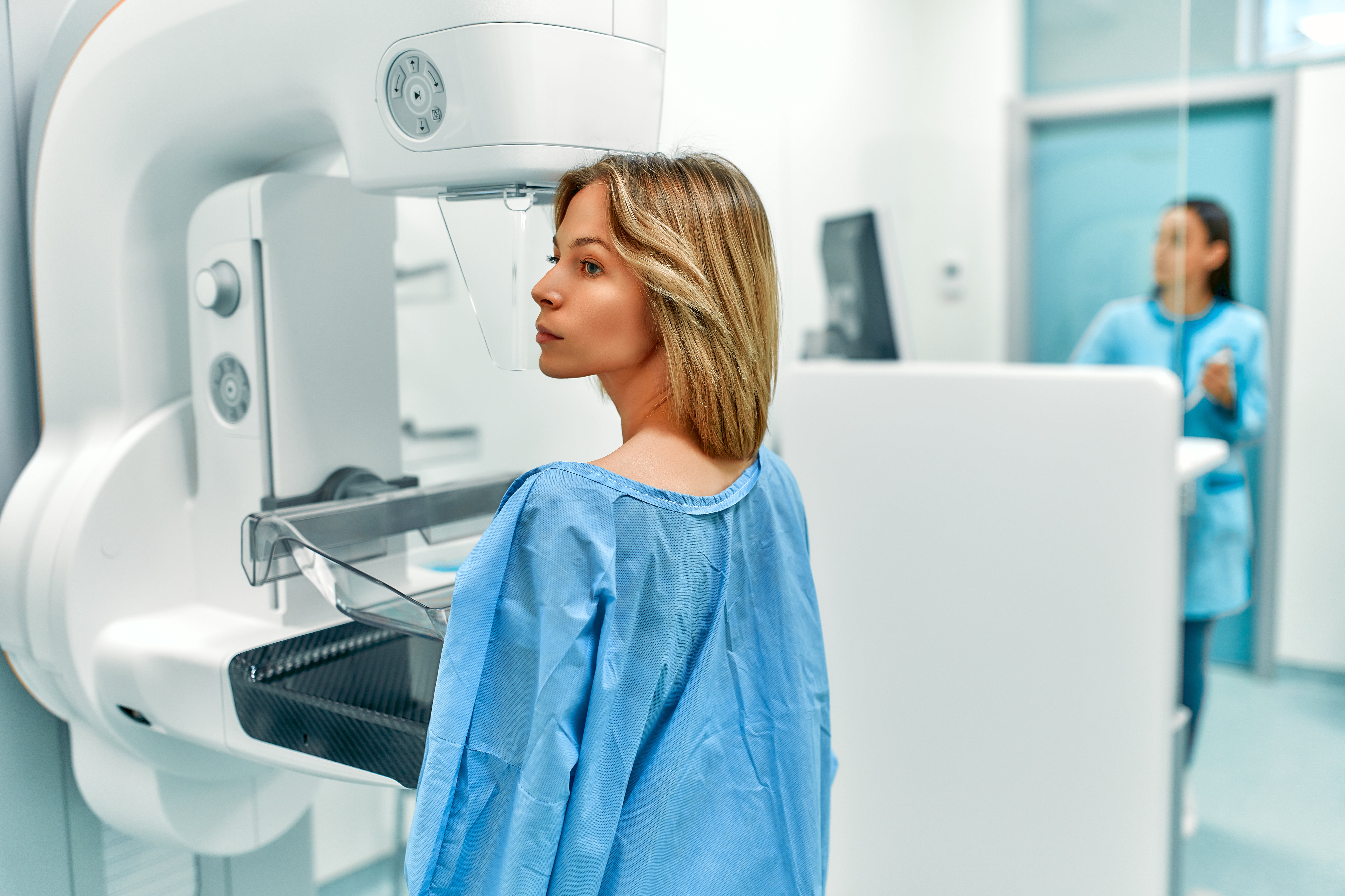
CEM uses updated standard mammography equipment to obtain low-energy and high-energy images following an injection of an iodine-based contrast. Subtraction images are then created that depict enhanced findings like those of contrast-enhanced MRIs, and the low-energy images are comparable to standard digital mammograms.
In a clinical study of 601 women at higher risk of breast cancer—87% of whom had dense breasts—led by Berg and funded by the Pennsylvania Breast Cancer Coalition, researchers added CEM to digital breast tomosynthesis (DBT), a 3D mammography technique, to evaluate its impact on incremental cancer detection rates, false positive recalls for additional testing, and the positive predictive value of biopsies performed. They found that adding CEM to DBT significantly increased early breast cancer detection but also increased false positives.
“Despite the false positives, CEM proved that it can catch very small tumors,” Berg said. “In the 12 women diagnosed with cancer in the study, we found six cancers that were only visible using CEM, which corresponds to an added cancer detection rate of 10 per 1,000. Five of those six cancers were invasive, with a median size of only 0.7 cm, and all were caught before spreading to lymph nodes. Importantly, there were no cancers found because of symptoms in the following year.”
These results are similar to those of prior studies where the cancers found on CEM were easily treated and have excellent prognosis, according to Berg.
In a separate, overlapping study, Berg found that about 70% of women who have had both examinations prefer CEM to MRI.
“Because mammography machines are widely available and most are easily upgraded to allow CEM, it is much easier to expand capacity for CEM than to install more MRI machines,” said Berg.
Currently, CEM is FDA-approved only for diagnostic use. Insurance typically covers the cost of the diagnostic mammogram, but copays and deductibles may apply, and there is typically an additional charge for the contrast. Berg indicates “we are just starting to offer CEM for patients with symptoms or newly diagnosed cancer at UPMC Magee-Womens Hospital, and we have direct CEM-guided biopsy capability as needed.”
The study, “Screening for Breast Cancer with Contrast-enhanced Mammography as an Alternative to MRI,” was published by the journal Radiology on June 17, 2025.
Media contact: HSNews@pitt.edu
